
Ultrasound to Measure Atherosclerosis
Atherosclerosis occurs on the inside of a vessel. The original BioImage study used a number of imaging techniques to find and quantify atherosclerosis. This included: by CT-scan, which uses X-ray radiation, MRI, which uses strong magnetic fields, PET, which uses radioactivity, and ultrasound, which uses soundwaves.
Of these four imaging techniques, ultrasound is the safest, lowest cost, and least burdensome. It is the only imaging method that, one day, has the potential to be recommended for routine screening. Ultrasound produces pictures of the inside of the body using sound waves. This is done with the use of a small transducer (probe) and ultrasound gel placed on the skin. The probe sends high-frequency sound waves through the gel into the body. The transducer collects the sounds that bounce back, and a computer then uses those sound waves to create an image.
Watch the video to see how the ultrasound is done.<
Carotid Ultrasound for Atherosclerosis Measurement from BioImage-2 on Vimeo.
Ultrasound has evolved over the past 30 years. Because of its safety, ultrasound is the preferred imaging method in obstetrics – imaging a baby within a pregnant woman. When we started the BioImage study, we partnered with the electronics company Philips, which is a leader in medical ultrasound equipment. Philips adapted an advanced probe that was commonly used in obstetrics to allow us to do an ultrasound of the large blood vessels in the neck.
A Surrogate Known as IMT
When we started the BioImage study, the most common method of ultrasound imaging of the arteries in the neck was measuring the thickness of a small portion of the carotid arteries. The carotid arteries can be found in the neck just below the skin and can easily be imaged with ultrasound. That thickness measurement is known as intima-media thickness, or IMT. IMT is measured by looking along the length of the vessel and by measuring the particular wall thickness just below where the artery splits. A thicker than normal IMT is not plaque, but was thought to be a measure of long-term blood vessel damage, and could be used as a measurement of risk.

Figure 1: IMT Measurement of Carotid Artery
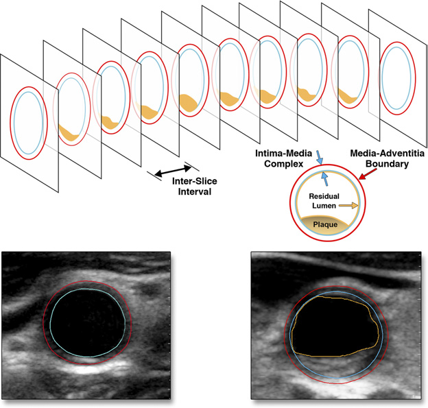
IMT is determined by measuring the distance between two parallel lines seen on images of the vessel wall of the common carotid artery (CCA) in a longitudinal view. These two lines represent layers of the vessel wall: from the wall that lines the vessel on the inside (intima), to the next boundary known as media-adventitia.
About 3D Ultrasound
In the BioImage study we measured IMT too, but, additionally, we looked for plaque along the entire length of the carotid arteries in the neck. We did this by turning the probe 90 degrees – instead of seeing the vessel as a long tube we were now looking at a slice. We then made a short ultrasound movie by moving the probe in about 10 seconds from the breastbone to the corner of the jawbone. This images the entire length of the carotid arteries (figure 3). At first, the ultrasound technicians did this by hand. Later in the study we also used a new motorized probe from Philips, where the ultrasound kept the probe in place and a small motor moved the transducer within the probe to provide a similar sweep. Because the sweep allows us to create a three-dimensional image of the arteries and the plaque lesions, we described this as 3-D ultrasound of the carotid arteries. This new way of 3-D ultrasound imaging of the carotid arteries produced striking results: of the 6,101 participants that had the new ultrasound imaging performed, 78% were found to have plaques in one or both carotid arteries. This was nearly double what others had reported, most likely due to our advanced way of imaging.


You must be logged in to post a comment.